
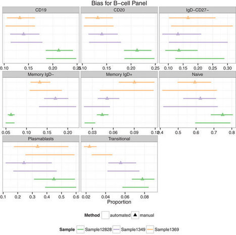

Standardization of immunophenotyping requires careful attention to reagents, sample handling, instrument setup, and data analysis, and is essential for successful cross-study and cross-center comparison of data.
Get Started for FREE
Sign up with Facebook Sign up with X
I don't have a Facebook or a X account


 Your new post is loading...
 Your new post is loading...
Your new post is loading...
Standardization of immunophenotyping requires careful attention to reagents, sample handling, instrument setup, and data analysis, and is essential for successful cross-study and cross-center comparison of data. 
No comment yet.
Sign up to comment

Developing cellular biomarkers is particularly challenging when it comes to biomarker validation, because flow cytometry has limitations regarding sample stability and multiplexing. Moreover, the development of highly multiplex flow cytometry assays is time-consuming and expanding existing panels can be quite cumbersome. ChipCytometry is a new, imaging based, cytometry technology bridging the gap between biomarker discovery and clinical biomarker validation through flexible “sequential singleple
Alfredo Corell's insight:
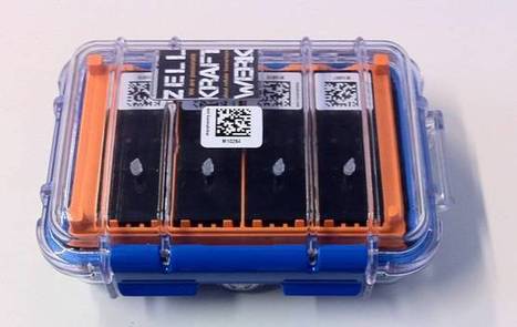
Some guidelines by Zellkraftwerk, the company developing the new called "chipCytometry"

An improved assay for assessing MRD status in patients with multiple myeloma is contributing to the momentum for using MRD as a surrogate endpoint for survival in clinical trials and as a tool with the potential to help guide therapy choices.
Alfredo Corell's insight:
In March, the International Myeloma Foundation (IMF) launched a new, automated flow cytometry test that is sensitive to 10-6 (1 cell in a million) and provides a computerized printout of MRD levels. The new test offers a more reliable method of assessing MRD status in an area that currently lacks standardization, said Durie, who is cofounder and chair of the IMF. - See more at: http://www.onclive.com/publications/oncology-live/2014/april-2014/new-mrd-assay-helps-move-testing-forward-in-multiple-myeloma-research/1#sthash.0RGCy54i.dpuf
Rapid Flow Cytometric Prenatal Diagnosis of Primary Immunodeficiency (PID) Disorders: http://t.co/FVhdPQz5vY #flowcytometry
Alfredo Corell's insight:
February 2014 Abstract Objectives Primary Immunodeficiency diseases (PID) are a heterogeneous group of inherited disorders of immune system. Immunophenotypic evaluation of PIDs using flowcytometry provides important clues for diagnosis of these disorders, though confirmation requires identification of underlying molecular defects. Prenatal diagnosis (PND) forms an important component of management in families affected with severe PID. However, molecular diagnostic facilities for each of these diseases are not available and may not be possible to perform in all cases. In such scenario we opted for phenotypic prenatal diagnosis by cordocentesis for families with index case having immunophenotypically well characterized PID. MethodsNormal reference ranges of lymphocyte subsets, CD 18/CD11 integrins on leukocytes, MHC class II expression and oxidative burst activity of fetal neutrophils at 18 weeks of gestation were previously established on 30 cord blood samples. PND was performed in 13 families with PIDs. Maternal contamination was ruled out by VNTR analysis. ResultsOut of 13 fetuses, nine were found to be unaffected (three cases with leukocyte adhesion deficiency (LAD-I), four cases with severe combined immunodeficiency diseases (SCID), one with X-linked agammaglobulinemia (XLA), and one with chronic granulomatous disease (CGD)] and three were found to be affected (one with T-B+NK-SCID, one with MHC class II deficiency and one with LAD-I). Diagnosis was confirmed by testing the cord blood samples after delivery and further follow-up of the children. In one family diagnosis could not be offered due to maternal contamination. No procedure related complications were observed. ConclusionFlowcytometry offers rapid and sensitive method for prenatal diagnosis and genetic counseling for selected phenotypically well characterized PID in cases where molecular diagnostic facilities are not available.
Alfredo Corell's insight:
Cytometry Part B: Clinical Cytometry - Volume 84, Issue 5 Pages 279–357 Issue edited by: Marie C. Béné, Gerald E. Marti
If you are looking to invest in Flow Cytometry technology, this guide aims to educate you on the essential information needed to assist your decisions.
Alfredo Corell's insight:
I've beem reading some parts of the document and I would rather expect a less tendentious results
A new dimension in cytometry and immunohistology
Alfredo Corell's insight:
Biomarker: 41-plex Panel Out Now! High-content human immune cell phenotyping based on Chipcytometry Technology The Human Immunecell Phenotyping Panel (HIPP) comprises: CD3, CD4, CD5, CD8, CD10, CD11b, CD11c, CD14, CD16, CD19, CD20, CD24, CD25, CD27, CD31, CD38, CD45, CD45RA, CD45RO, CD56, CD69, CD71, CD86, CD206, HLA-DR, IgD, IgG, IgA, T-bet, FoxP3, ROR gt, Helios, IL1b, IL8, IL10, IL12, IL17A, IL17F, IFNy, TNFa, pan Cytokeratin
Alfredo Corell's insight:
ESCCA 2013: INSPIRING AND INNOVATIVE The European Society for Clinical Cell Analysis (ESCCA) will organise ESCCA 2013 over 5 days in Luxembourg: the 13th Euroconference on Clinical Cell Analysis (12 - 14 November)the 9th European Course on Clinical Cytometry (15-16 November)These meetings are of major importance for education and scientific exchange in the fields of basic, translational and clinical applications in cytometry. They offer a unique opportunity to learn, to share information and scientific experience, to seek collaborations and to network - in short, to be involved and to meet old and new friends.
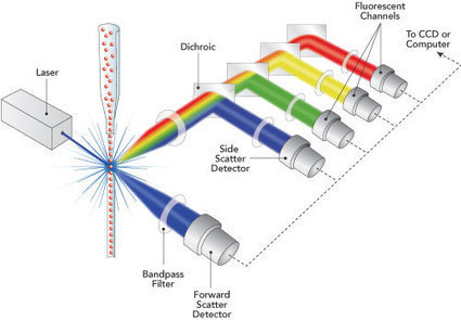
Create flow cytometry settings that allow for standardized consistent fluorescence intensity target values.
Alfredo Corell's insight:
Guidelines and steps to standarize Flow cytometry.
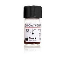
Streck Introduces Flow Cytometry Control for CD117 read Streck Laboratories Inc. news in the SelectScience scientific news archive
Alfredo Corell's insight:
CD check for abnormal CD117 expresión control
Visit our online shop: http://www.nitzipper.com nitzipper® is a new and revolutionary bioconjugation technology that allows to quickly, easily, and effective...
Alfredo Corell's insight:
Conjugate any particle or biomolecule with the conjugate you need for your research or diagnostic purpouses
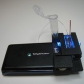
UCLA professor Dr. Aydogan Ozcan and his team have created a compact, low-cost fluorescent microscope and flow cytometer out of a cell phone camera.
Alfredo Corell's insight:
While you’re running around taking silly photos with your smartphone of what you ate for lunch today, a researcher at the University of California, Los Angeles, has found a way to photograph microscopic cell samples using a cell phone camera. Dr. Aydogan Ozcan, a professor and head of the Ozcan Research Group at UCLA, and his team of electrical scientists and bioengineers have developed a device that fits onto any camera-equipped cell phone that could benefit doctors and scientists in countries with limited clinical supplies. The research findings were recently published in the Journal of Visualized Experiments(JoVE, subscription required). Read more: http://www.digitaltrends.com/photography/ucla-scientists-turn-lowly-cell-phone-camera-into-fully-functional-research-microscope/#ixzz2QooMoN2V Follow us: @digitaltrends on Twitter | digitaltrendsftw on Facebook
Culturing of human retinal pigment epithelial cells (hRPE) is the initial step in cell therapy of some retinal diseases. To transfer these cells into clinical use, it is necessary to guarantee that they are well differentiated and contamination free. Fluorescence microscopy is the easiest method to do this, but it is associated with operator subjectivity, and the results are highly variable. The aim of this study was to demonstrate the practicality of implementing flow cytometry (FC) analysis to determine the purity of human RPE primary cell cultures. An ARPE19 cell line, human skin fibroblasts, hRPE, and human corneal epithelial cells were analysed by FC to determine the percentage of the hRPE population expressing RPE65 and epithelial and fibroblast proteins. The cell viability and DNA content also were determined. FC analysis showed that the hRPE cells were healthy, stable, and expressed RPE65 protein in the study working conditions. The density of RPE65 protein expression decreased during passages 2 to 10, which was confirmed using a Western blot technique. However, the hRPE cells did not express the 112-kDa epithelial and fibroblast proteins in the current working conditions. These findings suggested that FC facilitates a detailed analysis of human RPE primary cell cultures, a necessary step in developing new cell therapies for retinal diseases.
Alfredo Corell's insight:
Journal of Immunological Methods Volume 389, Issues 1–2, 29 March 2013, Pages 61–68 Flow cytometry assessment of the purity of human retinal pigment epithelial primary cell culturesGirish K. Srivastavaa, b, d, , 1, ,Roberto Reinosoa, b, 1,Amar K. Singha,Ivan Fernandez-Buenoa, b, d,Mario Martinoa, b,Maria T. Garcia-Gutierreza, b,J. Carlos Pastora, d,Alfredo Corella, b, c
|
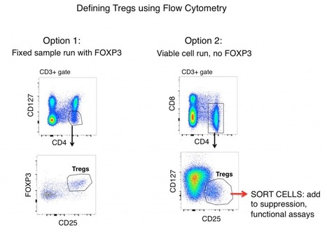
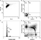
T-reg or T-regulatory cells have direct roles in both autoimmunity and responses to pathogens. Here we'll show you how to quantify Tregs.
Alfredo Corell's insight:
Gating Strategies For Defining Tregs By Flow Cytometry
The standard Treg gating strategy for both mouse and human samples (after first gating out doublets and gating on live cells) includes the antigens CD3, CD4, CD25, FOXP3, and CD127. When looking solely at antigen expression, Tregs are often defined as CD3+, CD4+, CD25hi, FOXP3+, and CD127lo (shown in the figure below as Option 1). Using these markers, a clear population is often visible from samples such as mouse splenocytes and human PBMC.
Alfredo Corell's insight:
Educational Objectives
This two day course will cover the fundamentals of flow cytometry to provide a deeper understanding of the critical concepts in flow cytometry. Through lectures and hands-on activities, students will learn core concepts in experimental design, implementation, staining, compensation, controls, statistical analysis, and troubleshooting. At the end of this course, students will have a better understanding of the complexities of flow cytometry and nuances to consider for their workflow.
Alfredo Corell's insight:
CYTO U and ISAC present their first online course: Flow Cytometry Biosafety
Authored by Kevin Holmes, Peter Lopez, Steve Perfetto, Hank Pletcher, and Ingrid Schmid, the Flow Cytometry Biosafety Course provides a summary of biosafety principles as they apply to flow cytometry and cell sorting. Register: http://cytou.peachnewmedia.com

Article first published online: 20 DEC 2013 DOI: 10.1002/cyto.a.22429 © 2013 International Society for Advancement of Cytometry
Alfredo Corell's insight:
This issue of Cytometry contains three reviews that further celebrate the longstanding, intense, and happy marriage between Cytometry and Immunology, as highlighted recently during the XV International Congress of Immunology, held in Milan, Italy, August 22–27, 2013. Scanning the scientific contributions of the Conference reveals that cytometric technology was used in the vast majority of abstracts and presentations. In other words, very few scientists did not switch on one or more lasers! It is obvious, that nowadays very few studies in the field of Immunology are possible without techniques based upon single cell analysis.
4th Edition reviews “one of the most valuable of its genre and…addressed to a wide audience…. Written in such an attractive way, being both informative and stimulating” (Zbigniew Darzynkiewicz, Trends in Cell Biology)
Alfredo Corell's insight:
This reference explains the science and discusses the vast biomedical applications of quantitative analytical cytology using laser-activated detection and cell sorting. Now in its fourth edition, this text has been expanded to provide full coverage of the broad spectrum of applications in molecular biology and biotechnology today. New to this edition are chapters on automated analysis of array technologies, compensation, high-speed sorting, reporter molecules, and multiplex and apoptosis assays, along with fully updated and revised references and a list of suppliers.
Alfredo Corell's insight:
Cytometry Part B: Clinical CytometrySpecial Issue: Validation of Cell-Based Fluorescence Assays: Practice Guidelines from the International Council for Standardization of Haematology and the International Clinical Cytometry SocietySeptember/October 2013
Volume 84, Issue 5 Pages 279–357 Issue edited by: Marie C. Béné, Gerald E. Marti

Flow cytometry applications, click for technical information and a full range of flow cytometry (FC) related products. FAST delivery, expert technical support.
Alfredo Corell's insight:
Some tips, protocols and applications of flow cytometry

Genetic profiling to identify specific cell surface markers for a highly suppressive Treg population.
Alfredo Corell's insight:
Tr1 cells uniquely expressed genes for two cell surface markers, CD49b and LAG-3.
Alfredo Corell's insight:
Registration is in full swing and space is limited. The tentative registration deadline is May 17th. In order to guarantee your spot please complete your registration!
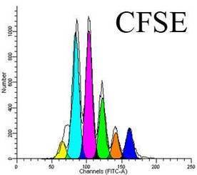
Measuring proliferation of lymphocytes using CFSE is not a foolproof protocol.
Alfredo Corell's insight:
A simple and useful review about This cytometry-based technique to asses lymphocyte proliferation
Alfredo Corell's insight:
ESCCA Summer Schools represent our new policy towards the development of residential, full-immersion courses which will pave the way to the specific certification of technical and academical operators, widely felt as a fundamental step for laboratory accreditation. Part of the courses will be organized nationally, in local languages and in full collaboration with the local cytometric society. An International course will be also organized in English, for the benefit of the operators from smaller Countries that cannot enjoy the support of a local society.
|


This study follows the “open science” trend by providing complete transparency of data and results, ensuring that reproducibility can be verified21,22,26. All materials, including primary data files, processed data, workspaces and analysis code, are made freely available using existing data standards and providing a valuable resource to the experimental and computational communities.